Illustration Techniques Assignment 1 Objective: Produce a fully rendered, highly detailed anatomical line illustration done in Corel Painter 11 for publication in college textbook.

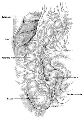
Anatomical Visualization Assignment 4 Objective: Produce an anatomical diagram identifying the location of thoracic or abdominal organs relative to surface landmarks and skeletal anatomy. Process: A human torso in anatomical position including all relevant surface anatomical landmarks was drafted using multiple anatomy atlases by Gilroy, Rohen, Clemente, and Moses. On an overlay-tracing sheet, the deep skeletal anatomy was drafted in registration with the surface anatomy using the same anatomy atlases with the addition of cadavers and skeletons from the gross […]
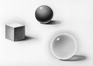
Third assignment in Anatomical Visualization. Part 1: Render basic forms with an upper-left light source. Posterior View of Anomalous Inferior Vena Cava with Azygos Continuation Part 2: Illustrate one of two case reports of anomalous inferior vena cava with azygos continuation with emphasis on the volumetric illusion of form through the use of light and shade. I chose to illustrate Anomalous inferior vena cave with azygos continuation in a Japanese man by Tohno. Process: Other articles concerning similar anomalies were […]
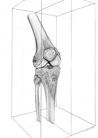
Second assignment in Anatomical Visualization. ¾ anterolateral view of the right knee joint in two-point perspective. After all my bad talk about this assignment, I soon sprained my knee during a mugging. Good news is that I may have free digital copies of my knee X-rays soon!
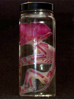
This fetal monkey with stained cartilage belongs to David Fisher, a medical illustrator employed at the University of Alabama at Birmingham, my undergraduate institution. David was the first medical illustrator I encountered and has heavily influenced and reaffirmed my pursuit of medical illustration.

First assignment in Anatomical Visualization taught by John Daughetry. Objective: Produce an accurate pencil sketch of an anatomical specimen that includes the bones of the shoulder region: clavicle, scapula, and proximal humerus. Illustrate the three bones and their articulations in an anterior and a posterior view. Then add the following anatomy to the bone drawing: Anterior view – coracoacromial lig., acromioclavicular lig., and coracoclavicular lig. Posterior view – supraspinatus m., infraspinatus m., and teres minor m. Process: Preliminary sketches were developed by transferring bone […]